  |
| HOME | THIS ISSUE | CALENDAR | GRANTS | BACK ISSUES | < BACK | NEXT > |

Patients, physicians, benefit from improved serviceDiagnostic imaging goes digitalby Maureen McGuire - September 5, 2006 |
||||
|
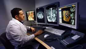
The Health Center's Department of Diagnostic Imaging and Therapeutics is now 100-percent digital, making services easier for patients and physicians alike. "Going completely digital is one in a series of major advances for our imaging services," says Anthony J. Borda, associate vice president for clinical operations.
"The days of cumbersome x-rays and large envelopes are over. Patients can leave appointments, or the Emergency Department or Urgent Care Center, with a CD of their test results. And physicians benefit from the fact that they can receive immediate, electronic copies of images." As soon as an image is taken, it goes into the Picture Archiving and Communicating System. A radiologist can then read the image anywhere there is computer access, within moments of the test. Imaging services are central to a wide variety of services offered throughout the Health Center. "Imaging is used for routine, preventive tests, as well as detailed state-of-the-art tests to detect the presence of heart disease by seeing actual blockages in arteries or the presence of tumors that can only be found through three-dimensional views of an organ," he says. Some of the most significant additions to the Health Center's imaging equipment in recent months include digital mammography, the PET/CT scanner, and open MRI. "Advances in imaging have brought about revolutionary changes in the way physicians can detect diseases and make life-saving early diagnoses," Borda says. "And the technology just keeps getting more sophisticated." Based on a study released by the National Cancer Institute in 2005, digital mammography is recommended for women under age 50 and women with dense breasts. Digital mammography is now available in Farmington; conventional mammography is available in East Hartford. "Mammography has been shown to save lives due to the early detection of breast cancer," says Dr. Joseph Walsh, the lead physician with UConn's general Ob/Gyn practice. The PET/CT scanner allows physicians to see a three-dimensional view of an organ and the location of a growth or tumor within the organ. It is a critical diagnostic tool for Health Center cancer specialists. It combines the technology of PET imaging - positron emission tomography - that allows experts to measure the metabolism, or chemistry, of organs and other tissues with CT scanning - computerized tomography - that shows a slice or cross-section of the body. The Health Center's open MRI allows for faster scanning, more detailed images, and more accurate diagnoses. It is located in the imaging suite of the Medical Arts and Research Building, which is home to the Health Center's Musculoskeletal Institute. "The MRI is open on three sides, so it doesn't feel like being put inside a big round doughnut," Borda says. "In the past, open MRIs only offered low-strength magnets. We offer a Philips open MRI scanner that provides images with the same excellent resolution as the closed MRI image, and the scan can be done much more quickly." The new imaging technologies complement the Health Center's existing technologies, including multi-detector, 16-slice, high-resolution CT scanners, and neuro-imaging. |
| ADVANCE HOME UCONN HOME |