For more archives, go to the Advance Archive/Search Page.
Protein Patterns In Muscles
At Heart Of Researcher's Work
Marie Cantino is a biophysicist, an associate professor of physiology and neurobiology, and director of the Electron Microscopy Laboratory. But when she looks at muscle filaments through the microscope, she sees patterns with the eye of an artist.
 |
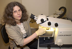
Marie Cantino, director of UConn's Electron Microscopy Laboratory, conducts research on how calcium regulates muscle contraction. Here she uses a special machine for cutting thin sections of frozen muscle tissue.
Photo by Melissa Arbo
|
"I really love looking at the images," she says. "At one point, I thought I might be an art major."
Analyzing the patterns - and determining how proteins in muscles are assembled and interact - is at the core of her work, which is funded by a $525,000 grant from the National Heart, Blood, and Lung Institute at the National Institutes of Health (NIH). She examines the muscles of bullfrog atria, or upper heart chambers, and skeletal muscle from rabbits through an electron microscope to find out how calcium regulates muscle contraction.
How the muscle proteins are arranged may yield clues to their function. For many years, according to Cantino, the importance of proteins known as actin and myosin, has been recognized. More recently other proteins, such as titin and C-protein, whose functions are not yet understood, are attracting interest as well. Mutations in their genes have been linked to a heart defect that can lead to sudden heart failure - the kind that sometimes occurs dramatically in athletes.
When part of a myosin molecule interacts with an actin molecule, a chemical reaction is catalyzed that produces force. Contraction in skeletal and cardiac muscles, which Cantino studies, is triggered by changes in the concentration of calcium inside the muscle.
It's been known for years that when a muscle is relaxed, calcium is present in low concentrations. When a nerve sends a signal to the muscle to go to work, calcium is released in the muscle cell. It binds to proteins in the muscle filament and allows myosin to bind to actin and produce force. The more myosin attaches, the easier it is for calcium to bind - an effect called "cooperativity, " which particularly interests Cantino.
The process is more complicated than scientists once thought. Cantino uses electron microscopes - instruments that use a beam of highly energetic electrons to examine objects on a very fine scale - to measure how calcium is distributed along the long filaments in contracting sarcomeres, the structures that the protein filaments form. Sarcomeres are the basic units that cause a striated muscle, such as skeletal or cardiac muscle, to contract, and they are what appear as 'stripes,' or striations, in muscle tissue viewed with a microscope. When a sarcomere shortens, it produces force.
Her technique of looking at muscle filaments a micrometer or so in length under an electron microscope is the only one that can directly measure the amount of calcium that is bound along the filaments, she says, and it is potentially important in deciding what's going on.
Calcium is important to cardiac muscle, Cantino notes. If you put a heart muscle into a calcium-free bath, it stops beating. But her interest is not in the clinical aspects of heart research, such as diet. She is more interested in the molecular mechanisms at work.
She first learned electron microscopy techniques in graduate school at the University of Wisconsin. When her adviser, a specialist in high voltage electron microscopy, moved to the University of Washington, she went too, and it was there that she obtained her Ph.D. in biophysics.
Her work at that time was on fertility. Cantino focused - with her eye for the aesthetics of materials - on patterns in sea urchin sperm.
"If you're working on this kind of thing, you have to be a person who's attracted to visual data," she says. She used to have to be in the darkroom a lot, developing pictures of what she had seen, but now she works on a computer, using Photoshop software - a tool familiar to graphic artists.
Since 1990, she has been director of the Electron Microscopy Laboratory administered by her department. The lab, where she works with academic assistants Jim Romanow and Steve Daniels, is now housed in the Biology/Physics Building. The facility has three transmission electron microscopes and one scanning electron microscope. It is kept impeccably clean and tidy.
"If a particle of dust gets in your sample, it looks like a redwood fell on it," Cantino says.
The preparation of biological samples for electron microscopy is one of her specialties. Biological materials can be 50 to 90 percent water, which will boil off in the vacuum that an electron microscope creates. She uses technically sophisticated methods to remove the water so the cells do not collapse, the sample is optimal, and the microscope is protected from damage.
Cantino teaches two courses in electron microscopy as well as the physiology half of BIO 107, an introductory biology course in which, she quips, students learn about the whole body in seven weeks. She also teaches muscle physiology to graduate students.
In addition, she collaborates with a physicist at Imperial College in London, where she has twice been a visiting researcher. Her work there looks at the structure of muscle filaments, trying to learn more about some of the proteins involved in defects that can lead to sudden heart failure. Her work has relevance in exercise physiology and issues of aging, she says, as well as better treatments for heart failure.

