For more archives, go to the Advance Archive/Search Page.
Chemistry Professor Unravels Mysteries Of Color Vision
Scientists have long known that birds, bees, and other creatures see the world in vastly different ways than do humans. But the mechanisms that allow fish to recognize colors, honeybees to navigate back to their hives, and birds to hunt in low-light conditions are little understood.
 |
|
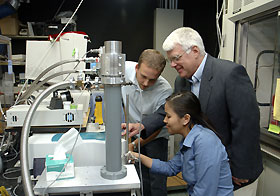
Robert Birge, professor of chemistry, with graduate students
Jerry Koscielecki, at back, and Lavoisier Ramos, at work in a
laboratory in the Chemistry Building. |
Professor Bob Birge and several graduate students are conducting research on intricate processes that occur within a specific protein in the rear of the human eye and in the eyes of some animals. Their findings are expected to contribute to a better understanding of the mechanisms and molecular details of color vision.
The Birge team is striving to understand events that unfold in rapid succession in certain cells in unimaginably tiny fractions of a second. The research would be daunting enough if the scientists were merely working with molecules that trigger changes in trillionths of a second, allowing color vision as we know it, but the challenge doesn't end there: As graduate student Lavoisier Ramos notes, "The proteins we work with are light sensitive, so we have to do our experiments in the dark, using only a red light such as you would find in a photography darkroom."
To appreciate the work being done by Birge and his students, it is necessary to understand the basic structure of the eye and the role of rods and cones in color vision.
Rods are photoreceptive — light receptive — cells in the retina of vertebrates. As rod cells are stimulated by light, they allow the eye to perceive the size, shape, and brightness of visual images, but not color. That function is handled by cone cells.
Within the rod cells is a protein called rhodopsin. It is this protein and the changes it causes that are at the heart of Birge's research. Rhodopsin, among other characteristi cs, permits vision under low light conditions.
Birge, the Harold S. Schwenk Sr. Distinguished Chair in Chemistry, hopes that a better understanding of the complex processes that lead to color vision will produce benefits for humanity. His research may facilitate the development of artificial retinas, for example, and improve understanding of night blindness and how Vitamin A contributes to vision, since a chemical within rhodopsin is derived from Vitamin A.
The nature of any creature's vision is determined by what light along the wavelength its eye is equipped to see. Humans, for example, can't see ultraviolet light, but honeybees and some birds can. And as any fly fisherman can attest, rainbow trout see in certain wavelengths better than humans do, allowing them to be very discriminating about precisely what color fly they will strike. As for bees, seeing ultraviolet makes pollen and nectar light up like a neon sign; and for birds such as kestrels, the scent marks of rodents become visible in ultraviolet light, enhancing hunting possibilities.
"UV cones help animals and insects find food and choose a mate," Birge says. "These are high-priority items and involve a level of competition with other creatures that demands optimal visual perception."
Humans, on the other hand, experience color based on combinations of red, green, and blue.
"Although humans have the genetic machinery to express UV cone pigments, evolution has simplified our color vision to be based on red, green, and blue cones," he says. "It is no coincidence that computer displays are based on RGB systems, because we can only perceive color based on red, green, and blue light.
Birge says this is not much of a shortcoming, however, "because the simple tri-color system can generate millions of different colors and we can distinguish color shading that approaches one part per million in content."
Collaborating with a team led by Professor Barry Knox at Upstate Medical Center in Syracuse, N.Y., the Birge group recently solved one of the key mysteries of UV vision: how a light-absorbing chemical called a chromophore went from having a neutral charge to acquiring a positive charge like other pigment chromophores. (The findings are published in the Proceedings of the National Academy of Sciences.)
Unraveling this mystery took two graduate student Ph.D. theses and an investigative tool called Fourier-transform infrared difference spectroscopy. Discovering how the chromophore picked up its charge — a proton — was like figuring out that a ping-pong ball has moved from one closet to a nearby one deep inside a 40-story building, Birge says.
To identify this particular "ping-pong ball," the group had to use the spectroscopic tool to determine that it had moved a few Angstroms, an Angstrom being a 10 billionth of a meter. The movement, tiny though it was, attracted the researchers' attention.
But the fact that the positively charged chromophore had shifted position infinitesimally posed a fresh problem for Birge's group. Knowing that visual pigment activation — the process that allows color and UV vision — cannot occur with an uncharged chromophore, the question became, "How did this chromophore acquire a charge?"
The group used a combination of spectroscopy and molecular biology to show that light itself is the trigger. After the chromophore absorbs light, it picks up a proton and thereby becomes charged. In the process, the UV cone now mimics a green cone in completing the activation of the nerve impulse, and allowing color vision to occur.

