For more archives, go to the Advance Archive/Search Page.
New Scanner At Health Center
Captures Detailed 3-D Images
In a matter of seconds, a new diagnostic scanning machine at the UConn Health Center can provide detailed, three-dimensional images of the most intricate anatomical structures and systems in the human body.
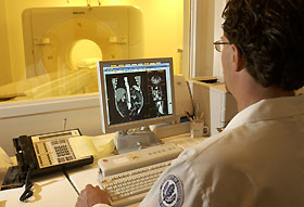 |
Jeff Moshka, a radiology technician, operates a new CT scanner at the Health Center.
The new equipment will improve diagnosis and treatment by providing accurate,
detailed images faster than previous technology.
|
By whittling away at the guesswork that often faces physicians in diagnosing and treating human disease, the machine continues the revolution in the practice of medicine that began when the technology was introduced in the 1970's.
"This machine provides extraordinarily accurate images in a fraction of the time needed for other tests," says Dr. Bipin Jagjivan, chief of diagnostic imaging. "It will change the way physicians work."
The Health Center bought and installed the new $1.6 million scanner during the summer, to replace an older machine.
CT scanners use radiation or X-rays to capture data from multiple angles. The information is then processed by a computer into an image on a video screen. Conventional CT scanners captured a single slice or image at a time. In the Health Center's new 16-slice spiral CT scanner, however, the X-ray beam rotates continuously, tracing a spiral path through the patient, taking multiple images and gathering continuous data with no gaps between images. The computer reconstructs the information into individual slices and combines them sequentially into a comprehensive image of the entire area scanned. Physicians can view the images almost instantaneously.
"It improves our ability to make accurate diagnosis of a variety of disorders, from cancer to cardiovascular disease and trauma," says Jagjivan. "The detail and clarity of the images allow for much more precise planning for surgery."
Blood flow to organs and limbs can be followed and imaged in three dimensions. This technique can be used for angiography, which visualizes blood flow in arteries throughout the body.
Typically, a catheter is inserted into an artery near the groin and snaked through the body. The 16-slice scanner eliminates the need for a catheter since the contrasting agent can be injected into a peripheral vein. The scanner can also perform virtual colonoscopy. Clear images of the inside of the colon can be obtained without inserting a colonoscope. By reducing the need for more invasive procedures, the chances for complications are lowered, Jagjivan says.
Dominic Romano, administrative director of diagnostic imaging, says the new technology is much quicker than conventional CT scanning. "The scanning time is cut by 50 percent for many tests, which makes it easier on patients. We used to have to ask them to hold their breath repeatedly so we could get a good scan or image. That's not really necessary for many procedures with this machine. It makes a big difference to elderly patients or to others in distress."

