|
This is an archived article.
For the latest news, go to the Advance
Homepage
For more archives, go to the Advance Archive/Search Page. |
||
|
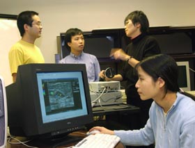
Zhu's Device Improves Breast
Cancer Detection
By Janice Palmer Each year, more than 200,000 women in the United States are diagnosed with breast cancer. Hundreds of thousands more experience a breast cancer scare because detection techniques are not always precise. About five years ago, it occurred to Quing Zhu, a physicist, that she might be able to develop a tool for making breast cancer detection easier and more accurate. Today, the device for which she holds a patent, is known as the "combined ultrasound and light imager", and it is undergoing clinical trials at the UConn Health Center in Farmington.
"We hope to improve diagnostic imaging of breast lesions by increasing detection sensitivity and specificity, which will save lives and reduce anxiety for patients," says Zhu, now an assistant professor of electrical and computer engineering at UConn. During the past several years, she has received three grants to develop the tool. Now, with a new $800,000 grant from the National Institutes of Health, she has a further four years to fine-tune it and prove its effectiveness. Her collaborators from the Health Center include Drs. Scott Kurtzman and Kristen Zarfos, breast cancer specialists, and Bipin Jagjivan and Mark Kane, radiologists. "This is a very exciting new tool that perhaps will significantly reduce the number of biopsies and operations women have to endure," says Dr. Kurtzman, who consults closely with Zhu on her research findings. He and Dr. Zarfos call in Zhu and her imager when they determine a patient would be an appropriate and willing test subject. When a breast mass is found, either by a mammogram or physical exam, breast cancer specialists often turn to ultrasound for determining whether the mass is a harmless cyst or solid lesion. But ultrasound is unable to accurately detect whether the solid lesions are benign or malignant, so a biopsy is often done. During the imager's clinical trials, Zhu conducts her test before a biopsy is undertaken. "For some time we've needed better tools to determine who needs a biopsy and who does not, " says Kurtzman, "because the majority of women who undergo biopsies are found not to have cancer. Think of the money saved. And, because results are much faster, women won't have to wait and worry about the biopsy results." The hand-held device Zhu developed combines ultrasound and near-infrare d optical techniques. Zhu credits her graduate students, especially Nan Guang Chen, her former postdoc, with developing the hardware and software needed for the imager. "I couldn't have done it without them," Zhu says. Near-infrared light is harmless, but highly sensitive to functional parameters that distinguish between benign and malignant lesions. While it can determine the type of lesion in the breast, it lacks spatial resolution, making it difficult to determine where the mass is specifically located. Ultrasound, on the other hand, emits high-frequency sound waves that bounce off tissues, producing a picture that pinpoints the lesion's exact location but fails to characterize the mass. Zhu is encouraged by the trials' early results, although another 200 or more patients are needed for conclusive results. She is also optimistic that the imager will lend a hand in the early detection process, because it is proving effective in detecting the smaller cancers, which often are the more aggressive types. But detection may not be the only role for the imager. Near-infrared light may also be effective in monitoring the success of anti-cancer drug treatment. Dr. Kurtzman explains that women with cancers too large to undergo lumpectomy are frequently treated with chemotherapy before the cancer is removed, in order to shrink the cancer and avoid a mastectomy. The hope is that the near-infrared light will tell doctors whether or not the cancer cells are responding to the treatment. Although Zhu's research has come a long way in just five years, she will not be satisfied with proving the imager's effectiveness. She describes herself as "a scientist devoted to the spirit of analyzing data and inventing new tools." All the information gathered during clinical trials will be used to create a database that will include data that might help the medical community better decipher the differences and similarities between the various types of breast cancers and aid in the accurate diagnosis of very early stage cancers. Zhu's ultimate goal, however, is "to contribute to the final eradication of this deadly disease. This is my dream," she says.
 |